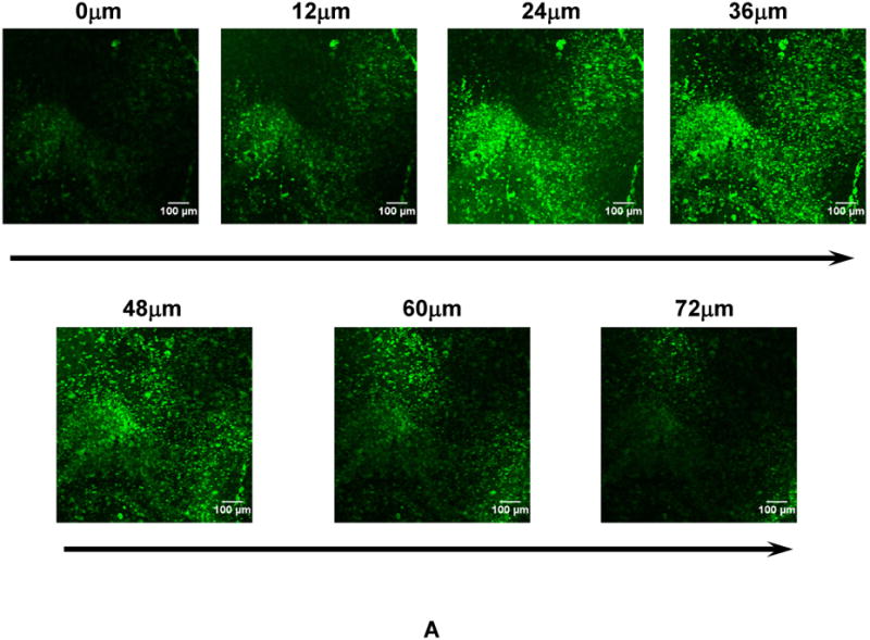
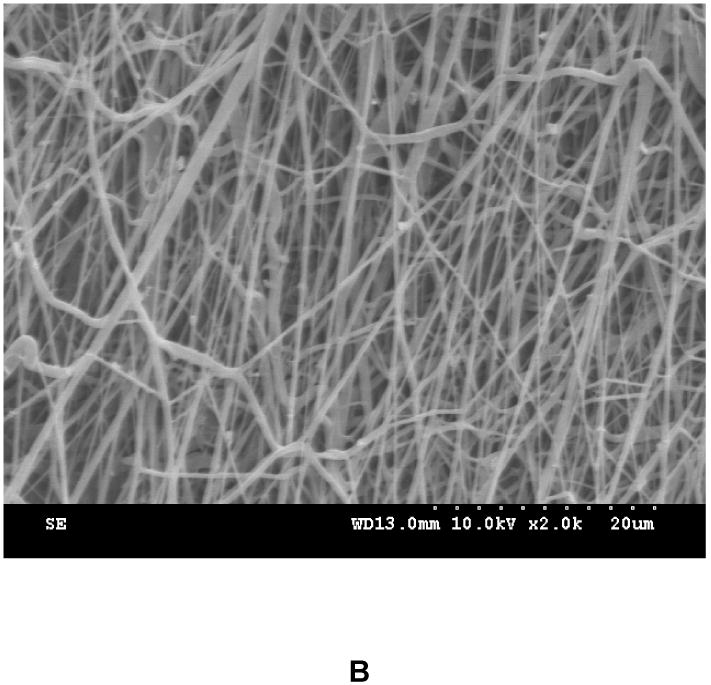
Figure 3.


(A) Z-stack confocal images of the tissue construct at different depth after one day of culture. Cells were stained with live cell stain CMFDA. (B) Surface topology of the tissue construct. The tissue construct was immersed in PBS for 1 day to completely remove gelatin and cells on the surface. This process allowed better imaging fibers, which otherwise can not be clearly imaged due to the interference of gelatin and cells.
