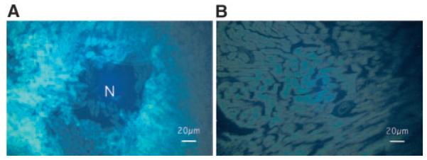
Figure 3.

Immunohistochemistry for HSV1-sr39TK (A) at the inoculated anterolateral wall and (B) at the nontransfected septum. HSV1-sr39TK is clearly stained with fluorescence (bright areas) in cardiomyocytes surrounding a needle track for the virus injection (shown as N) at the anterolateral wall, whereas little staining is noted at the septum.
