
Fig. 5.
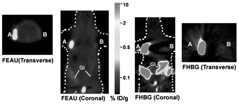
Micro-PET images of [18F]FEAU and [18F]FHBG uptake in a mouse xenograft model. One million of C6-sr39tk+ (A) and C6-tk+ (B) cells were implanted on the left and right shoulder of a nude mouse, and tumors were allowed to grow till they reach 3–5 mm of diameter. The mouse was first injected with ∼200 μCi of [18F]FEAU via tail vein and scanned in a micro-PET for 10 min after 1 h. Specific uptake of [18F] FEAU is seen in both tumors (A and B). After 24 h, the same mouse was injected with ∼200 μCi of [18F]FHBG via tail vein, and specific accumulation is again observed in both the tumors. Both transverse and coronal images of [18F]FHBG and [18F]FEAU scans are shown. [18F]FHBG showed relatively high signal in the gastro-intestinal tract (GI) because of clearance of the probe whereas [18F]FEAU exhibited very low signal in the gastro intestinal tract due to predominantly renal clearance. The color scale is in %ID/g tissue.