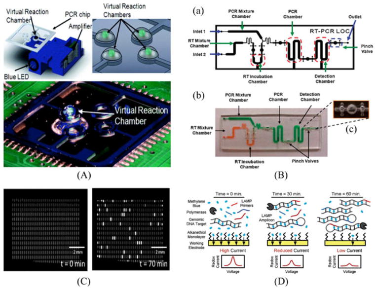
Fig. 5.
(A) Drawing of the on-chip PCR system with the optical configuration underneath. The integrated heater enables the light to interact with the sample placed above the heater. (Top right) A figure of four reactions at the glass on the top of the heaters. (Bottom) Photograph of the total system for PCR amplification. Reprinted from [57] with permission of RSC. (B) On-chip integration of reverse-transcription PCR for detection of pathogenic RNA species. Reprinted from [59] with permission of RSC. (C) Digitalized quantification of lambda DNA concentration by LAMP. An array of 535 wells is shown before (left) and after (right) incubation at 65 °C for 70 min. By counting the number of chambers with florescence, one can conjecture Reprinted from [68] with permission from RSC. (D) Microfluidic electrochemical detection via LAMP to detect pathogenic DNA for POC purposes. Reprinted from [69] with permission of Wiley.

