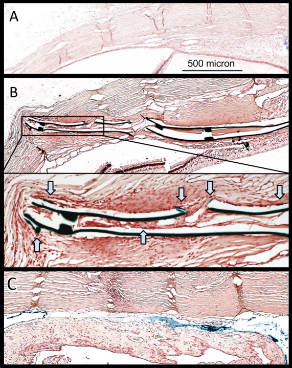
Figure 8.

Histology of implantation site. Light microscope images showing Prussian blue–stained slides. (A) Control eye. (B) GS-implanted eye shows blue tracer along the implant despite vessels in shunt lumen (inset, magnified view; blue arrows, tracer). Thin-walled, large vascular structures may resemble lymphatic vessels more than blood vessels. (C) PS surrounded by prominent fibrosis. The polypropylene dissolved in processing, while the space remains outlined by tracer.
