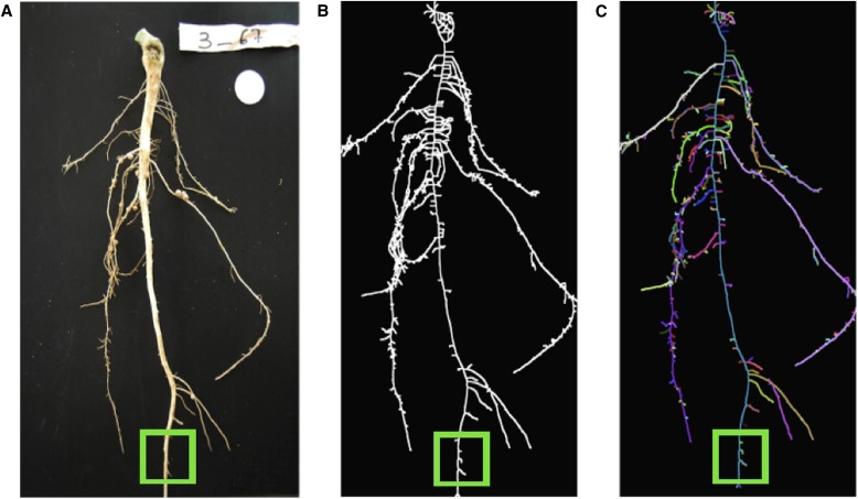
Fig. 8.
Detecting medial axis parts for removal. (A) A digital image of a cowpea root. (B) The medial axis derived from the image. (C) The segmented skeleton with random colors for each detected branch. Smaller branches are now removable by using a threshold and the known radius of the medial circle at the tip of each branch. The green box indicates an example area where additional branches can be removed.

