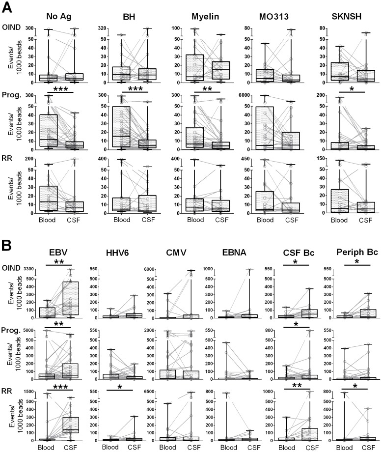
Figure 3. Enrichment of intrathecal T cell reactivities to foreign Ag's.
Intracellular cytokine secretion of each research subject was analyzed for IFN-γ+, TNF-α+ and double positive CD4+ T cell events. The sums of all cytokine positive events were normalized to beads. Paired T cell reactivities to unloaded DCs (No Ag) and auto-Ag's (A) and foreign Ag's (B) are shown for the peripheral (Blood) and intrathecal (CSF) compartment for each subject. Overlaid box plots represent median values with 25th and 75th percentiles; black lines indicate minimum and maximum values; *0.01<p<0.05, **0.001<p<0.01, ***p<0.001.

