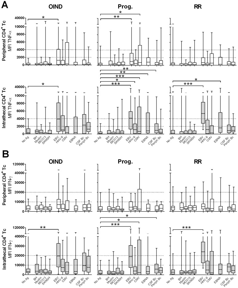
Figure 4. MFIs of peripheral and intrathecal CD4+ T cells.
MFIs of TNF-α- (A) and IFN-γ-producing (B) peripheral (upper panels) and intrathecal (lower panels) CD4+ T cells are shown for OIND, progressive (Prog.) and relapsing-remitting (RR) patients in response to all candidate Ag's. *0.01<p<0.05, **0.001<p<0.01, ***p<0.001.

