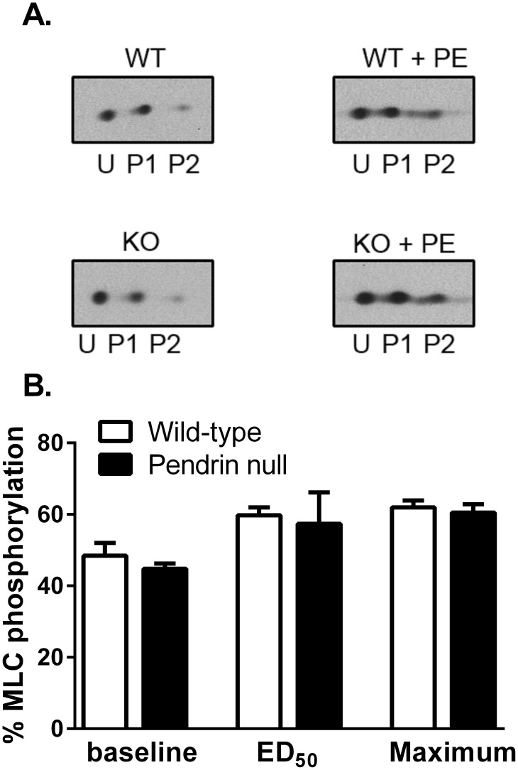
Figure 9. Pendrin gene ablation does not change the percentage of myosin light chain phosphorylation in the mouse aorta.
Aortas were isometrically mounted and phosphorylation was determined under either baseline conditions or following stimulation with phenylephrine at the ED50 concentration (0.3 µM) or at a concentration giving maximal stimulation (10 µM). The percentage of MLC20 phosphorylation was determined using 2-D electrophoresis. U represents the unphosphorylated light chain spot, whereas P1 & P2 are, respectively, the singly and doubly phosphorylated light chains. N = 4 in each group.

