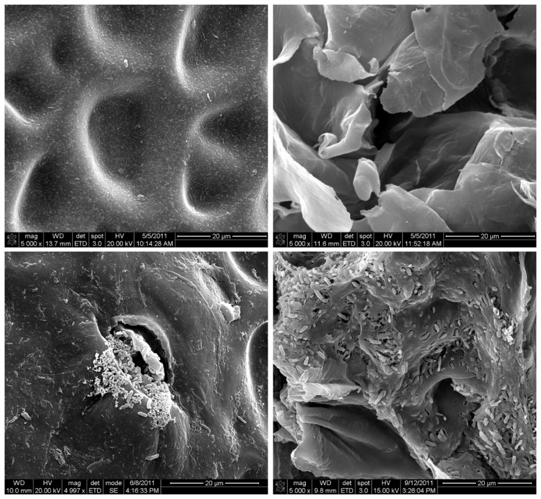
Figure 6. Scanning electron micrographs of cantaloupe rind surface at fruit maturity.

(A) Rind inoculated with 0.1% peptone water; (B) Crack on rind inoculated with 0.1% peptone water; (C) Rind inoculated with E. tracheiphila and had a watersoaked lesion with masses of bacteria seen near a trichome scar; and (D) Crack on rind inoculated with mixed S. enterica + E. tracheiphila that had a waterloaked lesion. All observations were made at 5,000×; scale bar shows 20 µm.
