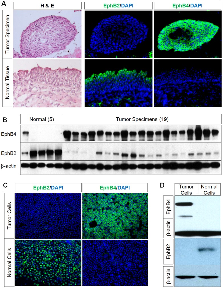
Figure 1. Reciprocal expression of EphB4 and EphB2 in bladder tumor and normal bladder cells.
A, Representative immunostaining on tumor and normal urothelium obtained from the same patient during cystectomy along with H&E stains showing EphB4 positivity in tumor specimens but not in normal tissue. In contrast there is EphB2 staining in normal tissue, but not in tumor specimens. Nuclei were counter-stained with DAPI. B, Western blot analysis of EphB4 and EphB2 in five normal bladder and 19 bladder tumor tissues shows high EphB4 expression in tumor but low or no expression in normal tissue and reciprocal expression pattern of EphB2. β-actin immunoblotting shows equal protein loading. C, Immunofluorescence staining of bladder cancer cell 5637 and normal urothelial cell PD07I also show strong EphB4 and absent EphB2 staining in tumor cells. There is strong EphB2 and absent EphB4 staining in normal cells. D, Western blot analysis shows EphB4 in tumor cell line 5637 but not in the normal cell line PD07I, whereas EphB2 is present in the normal cell line but not in the tumor cell line.

