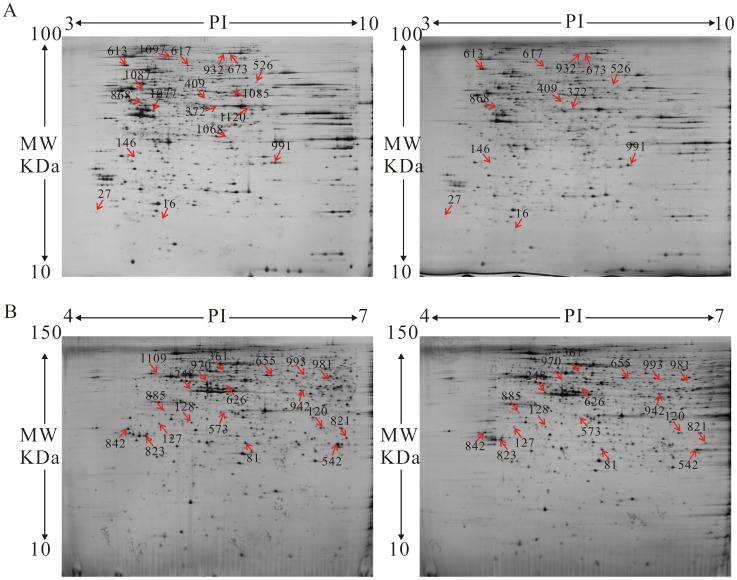
Figure 2. Proteomic analysis of BA-induced Hela cells was conducted by two-dimensional gel electrophoresis (2-DE).
(A) 2-DE was performed with 130 µg protein using 24-cm pH 3–10 NL IPG strips and 12.5% SDS–PAGE. (B) 2-DE was performed with 130 µg protein using 24-cm pH 4–7 NL IPG strips and 15% SDS–PAGE. The gels were visualized by silver staining and analyzed with Image Master Software.

