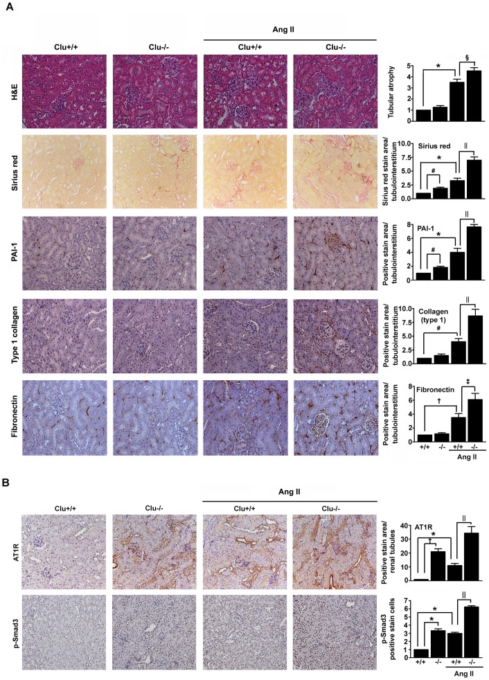
Figure 1. Effects of knockout of clusterin on Ang II-induced renal fibrosis.
Representative images of renal cortex sections from wild-type (Clu+/+) and Clu-/- mice treated with or without Ang II for 14 d. (A) The sections were stained with H&E or Sirius red, or were immunostained with antibodies targeting PAI-1, fibronectin, and type I collagen. The number of atrophic tubules was determined by measurement of the abnormal irregular and dilated tubular basement membranes in five random fields of H&E-stained sections under high-power magnification. Areas of positive staining with Sirius red and positive immunostaining with PAI-1, fibronectin, and type I collagen antibodies were quantified by computer-based morphometric analysis. (B) The sections were immunostained with an antibody targeting AT1R and p-Smad3. Areas of positive immunostaining were quantified by computer-based morphometric analysis. Data were normalized to the untreated wild-type and are expressed as the mean ± SEM of n = 5 random fields of each kidney (n = 5 in each group). * P<0.01, # P<0.05, and † P<0.001 compared with untreated wild-type mice and ‡ P<0.01, § P<0.05, and || P<0.001 compared with Ang II-treated wild-type mice. Original magnification, ×200.

