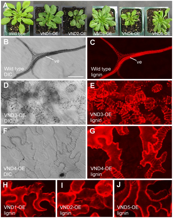
Figure 4. Ectopic deposition of lignified secondary walls in epidermal and mesophyll cells in leaves of VND1 to VND5 overexpressors.
(A) Three-week-old plants of the wild type and VND overexpressors (VND-OE). Note the upward curled leaves in the VND overexpressors. (B) and (C) Differential interference contrast (DIC) image (B) and lignin autofluorescence image (C) of a wild-type leaf. Note that lignified secondary walls were only present in veins (ve). (D) and (E) DIC image (D) and lignin autofluorescence image (E) of leaf mesophyll cells of VND3 overexpressors showing ectopic wall thickening and the corresponding lignin signal, respectively. (F) and (G) DIC image (F) and lignin autofluorescence image (G) of the leaf epidermis of VND4 overexpressors showing ectopic wall thickening and the corresponding lignin signal, respectively. (H) to (J) Ectopic lignin deposition in leaf epidermal (H and J) and mesophyll (I) cells in VND1 (H), VND2 (I) and VND5 (J) overexpressors. Bar in (B) = 29 µm for (B) to (G).

