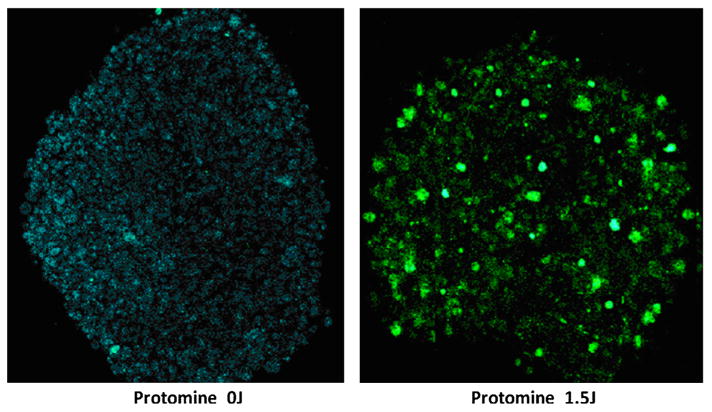
Fig. 7.

Two photon micrograph of GFP transfected U87 spheroids. PS/PTEN-GFP DNA concentration 4 μg/ml. a: The spheroids receiving no light showed only a faint auto fluorescence. b: PCI light treatment 1.5 J/cm2 showed greatly enhanced GFP production. Spheroid diameter 400 μm. Images were acquired at a depth of approximately 100 μm. The field of view for images was 600 × 600 μm2. [Color figure can be seen in the online version of this article, available at http://wileyonlinelibrary.com/journal/lsm]
