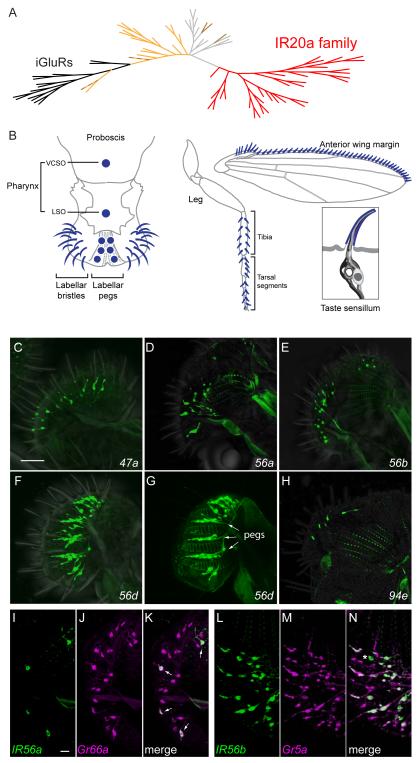
Figure 1.
The IR20a clade and members that are expressed in the labellum.
(A) Phylogenetic tree of IRs and iGluRs. The IR20a clade is represented in red and the iGluRs in black. IRs that are expressed in antenna are indicated in light orange (Silbering et al., 2011). Brown branches indicate IRs that have been found to be expressed only in taste sensilla; dashed brown branches indicate IRs expressed in both taste and olfactory sensilla (Croset et al., 2010; Zhang et al., 2013). The remaining IRs are represented in light gray. The alignment includes 33 IR20a clade members; IR56e and IR60f were omitted because they are short truncated polypeptides. See also Figure S1.
(B) Taste sensilla (dark blue) distributed in the pharynx (VCSO and LSO), labellum, leg and anterior wing margin. In addition, a typical taste sensillum is shown here with four different types of taste neurons (support cells are omitted for simplicity). Adapted from elsewhere (Dahanukar et al., 2007; Yarmolinsky et al., 2009).
(C-H) IR-GAL4 drivers that label cells in taste bristles (C-F, H, GFP) and taste pegs (G, GFP, arrows) on the labellum. (G) Labellum visualized at the level of the taste pegs, which lie between the pseudotrachea (autofluorescent ladder-like structures).
(I-K) IR56a-GAL4 (green, GFP) is expressed in a subset of Gr66a-RFP-expressing neurons (magenta) in the labellum. Small arrows indicate co-expression of both reporters.
(L-N) Almost all IR56b-GAL4-labeled (green, GFP) neurons are co-labeled by Gr5a-LexA (magenta, tandem-Tomato), except for a small proportion that is only labeled by IR56b-GAL4 (*). Images are z-projections of confocal images. Scale bars represent 40 μm in C-H and 10 μm in I-N. See also Table S1 and Figure S2.

