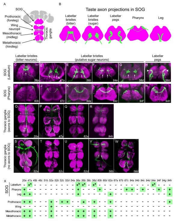
Figure 3.
IR20a clade GAL4 drivers label axons that target taste centers in the CNS.
(A) CNS of Drosophila, with regions contacted by taste axons highlighted in magenta. The SOG receives projections from the labellum, pharynx, and legs. The pro-, meso-, metathoracic and wing ganglia receive projections from the forelegs, midlegs, hindlegs and wing margins, respectively.
(B) Axonal projections (green) of taste neurons in the SOG. Axons lie in different planes along the z-axis: axons from labellar pegs and pharyngeal sensilla are anterior; axons from labellar bristles are in middle; axons from legs are posterior. Axons from the pharynx cross the midline in some cases (dotted line).
(C-H) Axons from labellar taste neurons. (C) Single arrow indicates axons of bitter neurons crossing the midline. (D-G, arrows) Axons are concentrated on both sides of midline, in a pattern resembling that of sugar neuron projections. In H, the arrowheads indicate axons that resemble those from labellar pegs. D includes projections from the legs.
(I-N) Axons from pharyngeal taste neurons. N includes projections from the legs.
(O-R) Axons that pass through the thoracic ganglia and project towards the SOG.
(S-W) Axons that terminate within the thoracic ganglia. Asterisks in S indicate projections into the wing neuropil labeled by IR52a-GAL4.
All images are from females. Z-projections of axons expressing GFP are in green and the synaptic marker Bruchpilot is in magenta. Scale bars in C and O represent 30 μm for C-N, and O-W, respectively.
(X) Summary of axonal projection patterns of neurons expressing GAL4 drivers. “b” and “s” indicate axons that resemble those of bitter and sugar neurons, respectively. IR56a-GAL4 labels axon projections consistent with its peripheral expression, in addition to other neurons (not shown).

