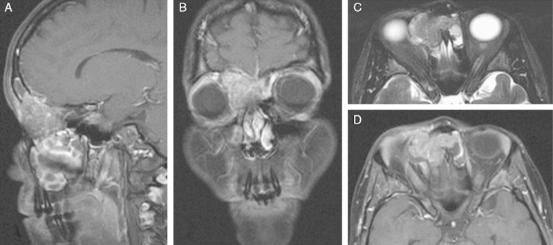
FIGURE 1.

Representative magnetic resonance tomography of case 1 showed a mass in the right anterior ethmoid cells extending into both frontal sinuses with destruction of the frontal bone and the right medial orbital wall. The lesion has a salt and pepper–like appearance on both postcontrast T1w images (A, B, D) and T2w images (C). Infiltration of the extraconal space and displacement of the right eye ball were noted.
