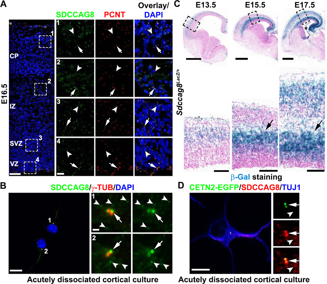
Figure 1. SDCCAG8 expression and localization in the developing cortex.
(A) Representative images of E16.5 cortices stained for SDCCAG8 (green) and PCNT (red), a centrosomal marker, and DAPI (blue). High magnification images of the representative areas (broken lines) in the CP (1), IZ (2), SVZ (3), and VZ (4) are shown to the right. Note the localization of SDCCAG8 at the centrosome (arrows) as well as the small puncta in the cytoplasm (arrowheads). Scale bars: 50 µm and 10 µm. (B) Representative images of two acutely dissociated culture cells (1 and 2) from E15.5 cortices stained for SDCCAG8 (green) and γ-TUB (red), another centrosomal marker. High magnification images of the centrosomal region are shown to the right. Note the localization of SDCCAG8 as two dots at the centrosome (arrows), as well as the cytoplasmic puncta (arrowheads). Scale bars: 10 µm and 1 µm. (C) Representative images of brain sections of Sdccag8Lacz/+ mice at E13.5, E15.5, and E17.5 stained for β-Gal (blue) and counter-stained with Nuclear Fast Red (magenta). High magnification images of the cortices (broken lines) are shown at the bottom. Note the selective elevation of Sdccag8 expression around the IZ (arrows), where newborn neurons reside prior to their radial migration to the CP. The asterisk indicates the expression of Sdccag8 in the choroid plexus. Scale bars: 500 µm and 100 µm. (D) Representative images of acutely dissociated cortical neurons expressing CETN2-EGFP (green) stained for SDCCAG8 (red) and TUJ1, an immature neuronal marker (blue). Note the localization of SDCCAG8 at the centrioles (arrows) and the cytoplasmic puncta (arrowheads). Scale bars: 10 µm and 2 µm.

