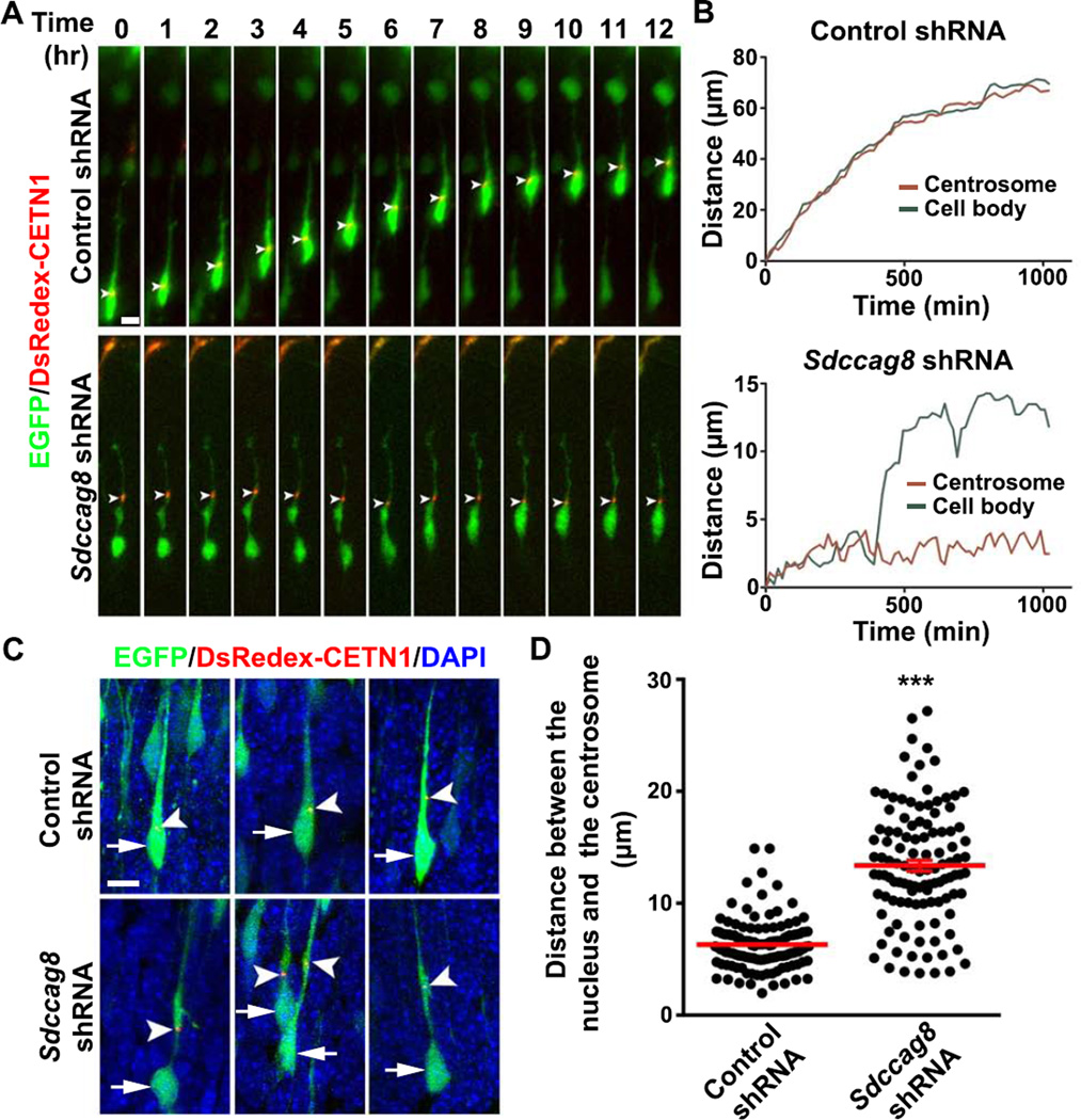
Figure 4. Depletion of SDCCAG8 impairs coordinated movement of the centrosome and the nucleus.
(A) Representative kymographs of bipolar migrating neurons expressing DsRedex-CETN1 (red) to label the centrosome and EGFP/Control (top) or Sdccag8 (bottom) shRNA. Arrowheads indicate the position of the centrosome. Scale bar: 5 µm. (B) Traces of the cell bodies (green) and the centrosomes (red) in migrating neurons in (A). (C) Representative images of bipolar neurons in E16.5 cortices electroporated with DsRedex-CETN1 (red) and EGFP/Control or Sdccag8 shRNA (green) at E13.5 and stained with DAPI (blue). Arrows indicate the cell bodies and arrowheads indicate the centrosomes labeled by DsRedex-CETN1. Scale bar: 10 µm. (D) Quantification of the distance between the centrosome and the nucleus (µm) in bipolar neurons. Each dot represents a neuron and red lines represent mean ± s.e.m. (Control, n = 131; Sdccag8, n = 114). ***, p<0.001.

