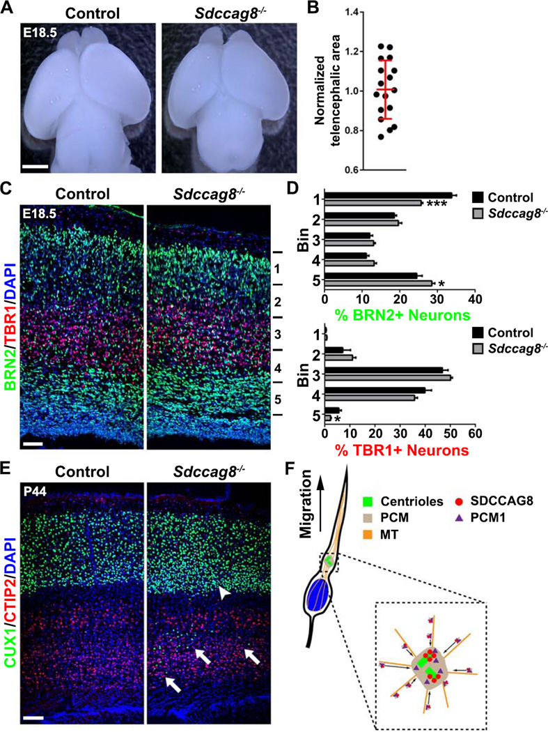
Figure 9. Sdccag8 knockout impairs neuronal migration in the cortex.
(A) Representative whole mount images of E18.5 Control (Sdccag8+/+ or Sdccag8+/−) and Sdccag8−/− brains. Scale bar: 2 mm. (B) Quantification of relative telencephalic area of Sdccag8−/− brains cortices to the littermate controls. Red lines represents mean ± s.e.m (n = 17). (C) Representative images of E18.5 Control (Sdccag8+/−) and Sdccag8−/− cortices stained for BRN2 (green) and TBR1 (red), two specific markers for the superficial and deep layer neurons, and with DAPI (blue). Scale bar: 50 µm. (D) Quantification of the percentage of BRN2+ and TBR1+ neurons in different bins of the cortex (Control, n = 7 brains; Sdccag8−/−, n = 6 brains). ***, p<0.001; *, p<0.05. (E) Images of P44 Control and Sdccag8−/− cortices stained for CUX1 (green) and CTIP2 (red), the respective superficial and deep layer neuronal markers, and with DAPI (blue). Note the presence of substantial CUX1+ neurons near the bottom of the cortex (arrows) and the relative high density of CUX1+ neurons at the bottom of the superficial layers (arrowhead), indicating a neuronal migration defect. Scale bar: 100 µm. (F) A model illustrating the role of SDCCAG8 in regulating PCM recruitment and neuronal migration in the developing cortex.

