
Fig. 2.
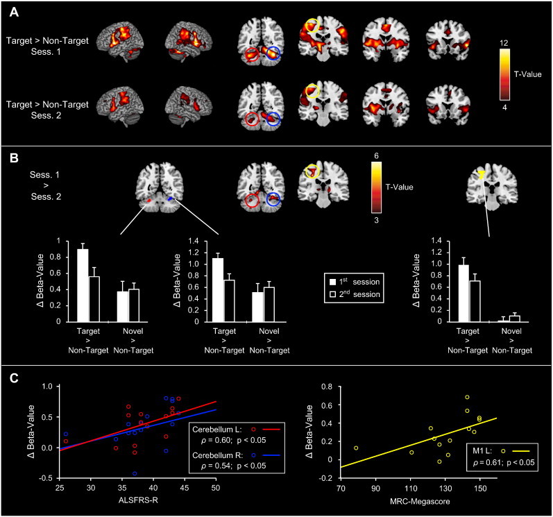
Functional changes in motor regions related to ALS disease-progression. A) Motor activity (“Targets > Non-Targets”) from the 1st and 2nd fMRI sessions. Motor-related activations were observed in the bilateral primary motor cortex, cerebellum, inferior frontal gyrus/ anterior insula, striatum, thalamus, and in the supplementary motor area. Note that maximum t-values and the extent of activations are considerably higher for the 1st than for the 2nd session. B) Direct comparison of motor-related activity across sessions. A decrease in motor-related activity from the 1st to the 2nd measurement was evident in the left primary motor cortex and bilateral cerebellum, which was confirmed in a ROI-analysis (see bar graphs). C) Correlation of ROI-results and the patients' clinical data. The decrease in motor-related activity correlated with the patients' ALSFRS-R scores for both cerebellar ROIs, while in the primary motor cortex it correlated with their MRC-Megascores.