
Fig. 3.
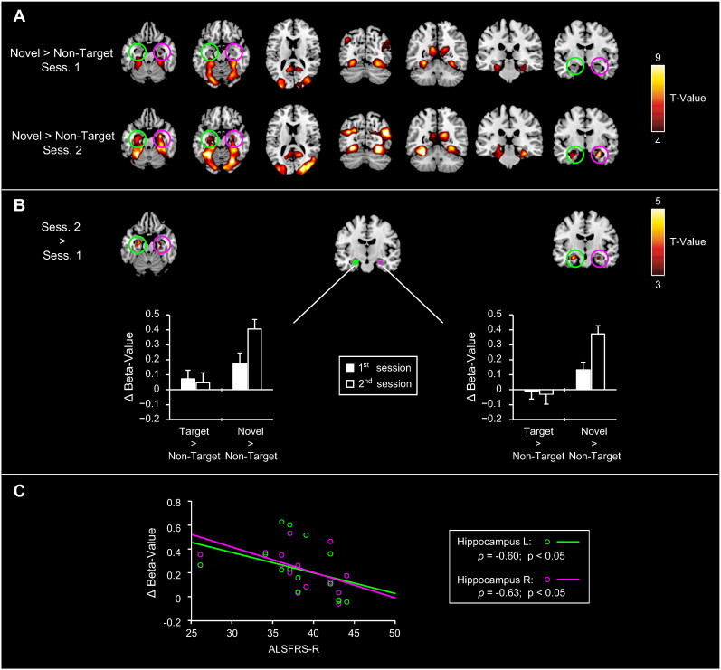
Functional alterations in novelty processing related to ALS disease-progression. A) Novelty-related activity (“Novels > Non-targets”) from the 1st and 2nd fMRI sessions. Novelty-related activity was observed across multiple ventral occipital and temporal regions, including the middle/superior occipital, fusiform, lingual and parahippocampal gyri. In addition, significant hippocampal novelty-related activations were evident during the 2nd measurement (compare activation maps between the upper and lower rows). B) Direct comparison of novelty-related activity across sessions. A decrease in hippocampal novelty-related activity occurred from the 1st to the 2nd measurement, which was confirmed by a subsequent ROI-analysis (see bar plots). C) Correlation of hippocampal ROI-results and the patients' clinical data. The hippocampal activation-increase showed a negative correlation with the patients' ALSFRS-R scores for both ROIs.