
Fig. 5.
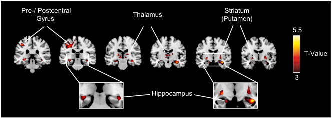
Regional atrophy in ALS patients in comparison to controls. Results of the between-group VBM analysis are superimposed on a standard MNI template thresholded at p < 0.001 (uncorrected). Clusters of significantly reduced MTR (ALS < controls) were detected within the bilateral precentral gyrus and several subcortical regions, including the bilateral hippocampus, thalamus and striatum (see Table 6 for MNI-coordinates and maximum t-values).