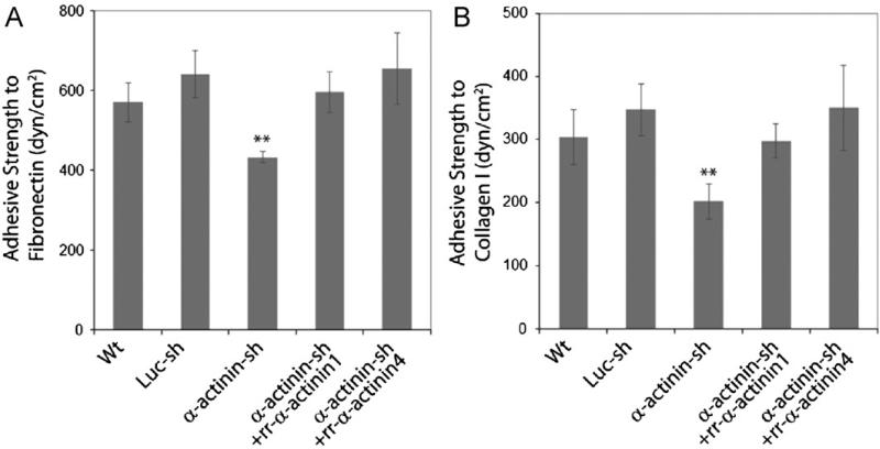
Fig. 5.
Cell adhesive strength to fibronectin and collagen type I is reduced in the absence of α-actinins. U2OS derived cells were plated onto fibronectin or collagen type I coated silicon substrate membranes overnight, initial images were captured, radial flow shear stress were applied, cells were then fixed and imaged. The critical radial position corresponding to 50% cell detachment was determined, adhesive strength was calculated. Data represent statistical result from three independent experiments.

