
Figure 6.
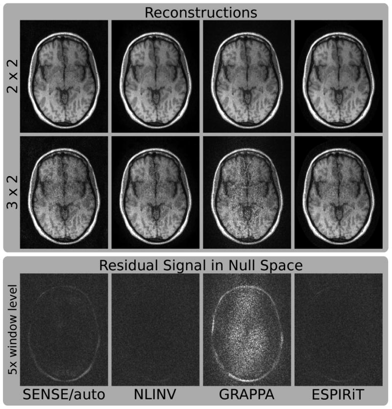
Images of a human brain. Fully-sampled data from an eight-channel coil has been retrospectively undersampled by factors of 2 × 2 and 3 × 2. Reconstruction has been performed using SENSE with autocalibration (SENSE/auto), nonlinear inversion (NLINV), GRAPPA, and ESPIRiT. The projection of fully-sampled individual coil images onto the null space has been computed for all methods and combined to a single image scaled by a factor of 5 (bottom row). For GRAPPA, the projection corresponds to a reconstruction operator corresponding to a regular 2 × 3 undersampling pattern. If the null space contains residual energy in addition to noise, this indicates errors in the calibration.