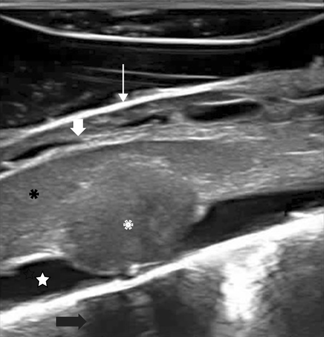
Fig. 2.

IoUS sagittal B-mode transdural imaging showing a dorsal intradural extramedullary tumor (neurinoma): ioUS scan identifies the tumor (asterisks) compressing the spinal cord (black asterisks), which appears slightly enlarged and hyperechoic due to spinal cord edema. Also the surrounding structures are depicted: dura mater (thin arrow), cerebrospinal fluid space (star), posterior rootlets (thick arrow) and the vertebral body (black arrow)
