
Fig. 3.
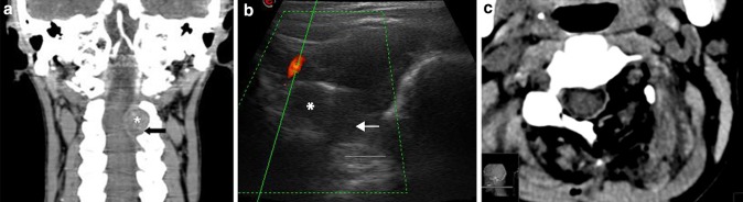
Extradural tumor: angio-CT coronal scan (a) showing a dumbbell extradural left C2–C3 neurinoma (asterisks) with bone scalloping (black arrow). IoUS B-mode imaging (b) with tumor (asterisks) evaluation also showing bone remodeling (arrow). Doppler imaging is used to evaluate vascular structures close to the lesion such as the vertebral artery. Postoperative CT scan (c) showing the complete tumor removal and bone work