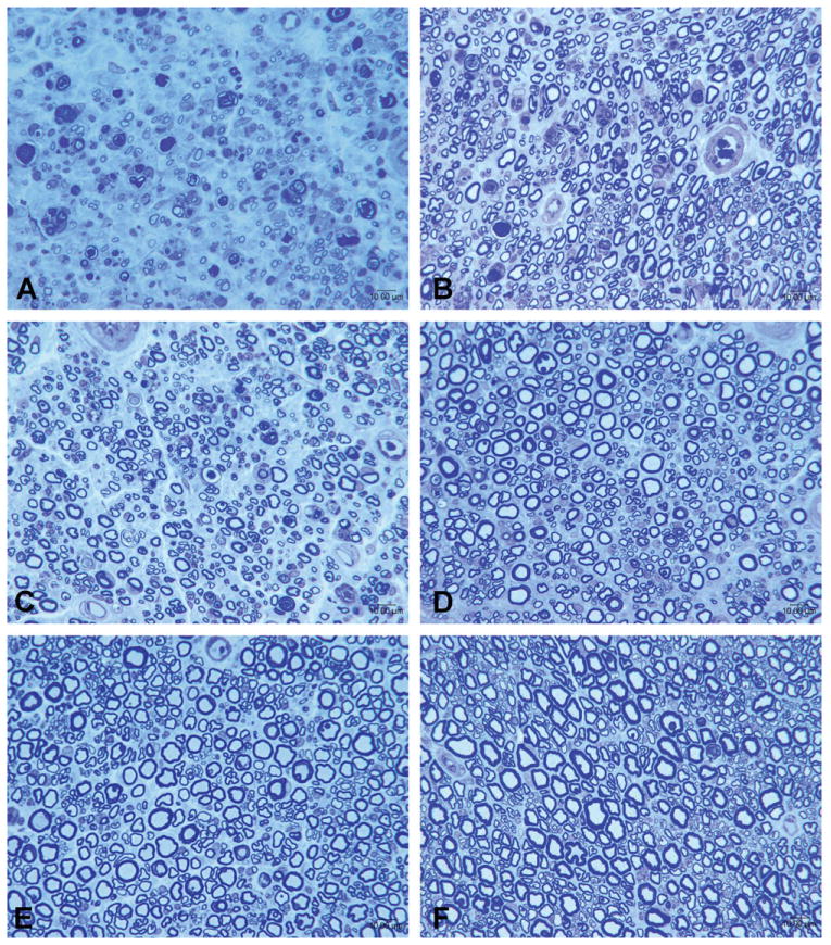
Figure 1.

Representative photomicrographs of sciatic nerves after end-to-end repair groups in animals sacrificed at serial time points. A: 1 month; B: 3 months; C: 6 months; D: 9 months; E: 12 months; F: 24 months. [Color figure can be viewed in the online issue, which is available at wileyonlinelibrary.com.]
