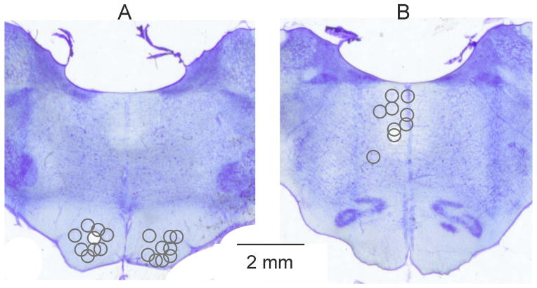
Figure 1. Sites of brainstem stimulation.
Reconstructions of the locations of the stimulating electrodes in the pyramidal tracts (A) and in and lateral to the medial longitudinal fascicle (B). The stimulation sites defined by electrolytic lesions made at the end of the experiments are indicated on representative sections of the medulla at the levels of the superior (A) and inferior (B) olives respectively. The circles in (A) and (B) correspond to the centres of the lesions and their diameters to the distances of estimated spread of current, within 0.2–0.3 mm from the electrode tip for 20–30 μA (see Fig. 11 in Gustafsson & Jankowska, 1976).

