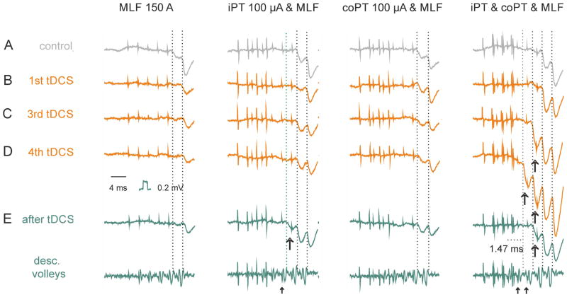
Figure 6. Examples of facilitation of IPSPs evoked from MLF during and after tDCS.
Records from a single GS motoneuron held through tDCS application. (A), control records before tDCS by various combinations of the stimuli as indicated. Note facilitation of IPSPs evoked by the last two MLF stimuli by either co or i PT stimulated separately or jointly. (B–E), records of IPSPs evoked by the same combinations of stimuli during the 1st, 3rdth and 4th periods of tDCS and 10 min after the last tDCS as indicated. Note facilitation of IPSPs evoked by not only the last two MLF stimuli but also an earlier stimulus after the 3th tDCS and two earlier stimuli during the 4th tDCS. Large arrows indicate earliest IPSPs facilitated by iPT. Small arrows indicate MLF volleys followed by these IPSPs. Vertical dotted lines indicate onset of IPSPs evoked by MLF stimuli. Other indications are as in Fig. 2. The sites of recording and stimulation and the underlying convergence of iPT, co PT and MLF fibres on reticulospinal neurons were as indicated in the diagram in Fig. 6.

