
Figure 3.
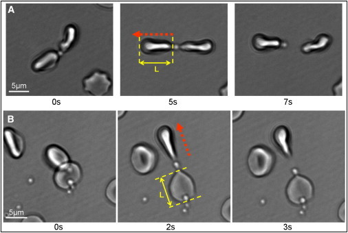
Selected frames taken from Movie S2 (A) and Movie S3 (B), showing optical tweezers-induced detachment of a postviable merozoite adhered to two erythrocytes. (Red arrows) Optical tweezers-directed movement. The cell-merozoite-cell chains are shown in a relaxed state, upon stretching in one direction, and upon detachment of the merozoite from one of the erythrocytes. Both erythrocytes are held in optical traps in panel A. In panel B, one of the erythrocytes is fortuitously attached to the cover glass. L is the cell length, and L = L0 + ΔL, where ΔL is the elongation and L0 is the cell length in the relaxed state. The detachment force F = κΔL, where κ is the cell stiffness used from Yoon et al. (11). To see this figure in color, go online.