
Figure 3.
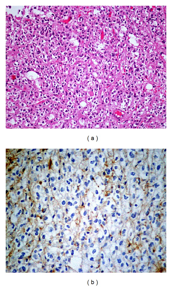
Histological examination. (a) Hematoxylin/Eosin coloration, with ×20 magnification, reveals moderately cellular neoplasm, consisting of cells with round nuclei and perinuclear light halo. (b) GFAP coloration, with ×40 magnification, reveals focal immunoreactivity of neoplastic cells (GFAP positive, CD68 negative cells).