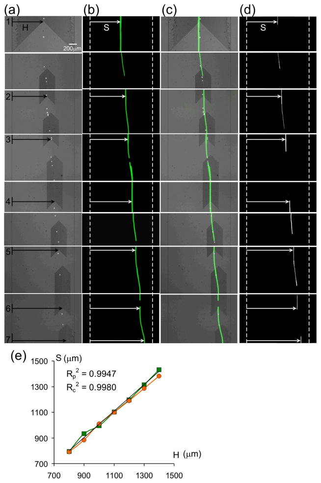
Figure 3.
Microvortex particle-guideing. (a) Bright-field microscopy, (b) fluorescent trajectories and (c) their superpositioned images of polystyrene beads focused and guided by locally patterned herringbone grooves in a microchannel. (d) Fluorescent trajectories of guided H1650 cells in the same device. (e) The lateral positions of the bead and cell trajectories S closely overlap with the lateral positions of the herringbone grooves H (the green line and red line represent polystyrene beads and H1650 cells respectively. The Reynolds number ~ 0.012 for both bead and cell guiding experiments.

