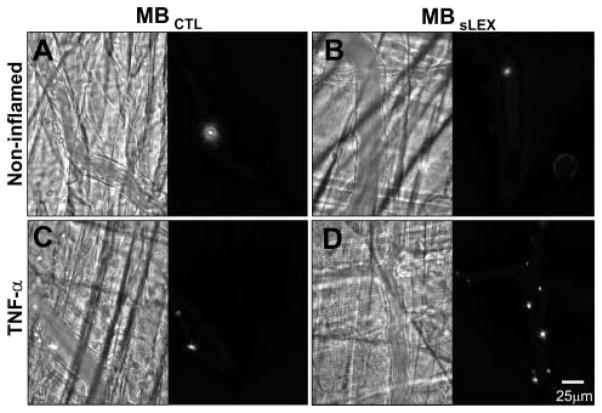
Figure 1.
Photomicrographs of murine cremaster microcirculation under basal conditions (A, B) or tumor necrosis factor–α–induced inflammation (C, D) after intravenous fluorescent MBCTL (A, C) or selectin-targeted MBsLex (B, D). Bright-field images are on the left and corresponding fluorescent images are on the right of each panel. There is greatest microbubble adhesion when MBsLex is injected into tumor necrosis factor–α–stimulated mice (D).

