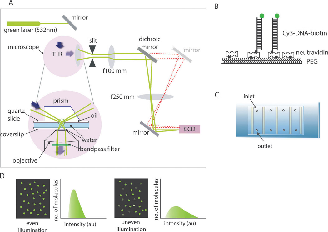
Fig. 1.
(A) Schematic diagram of the prism-type total internal fluorescence microscope (TIRFM) setup. (B) Fluorescently labeled and biotinylated-DNA immobilized to a polymer (PEG)-coated surface via biotin–NeutrAvidin interactions. (C) Slide assembly with flow chambers constructed for single molecule imaging. (D) Examples of even and uneven laser illumination of the single molecule surface.

