
Figure 2. CCL3 inhibits OB mineralization.
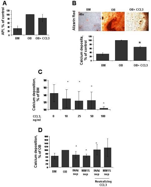
(A) The ratio of alkaline phosphatase activity relative to the amount of viable cells (API) in 2-week differentiated OBs with or without CCL3 50 ng/ml. (B) (upper panel) Representative image of alizarin red staining to detect mineralization from OB exposed to CCL3 for 3 weeks. (Lower panel) Quantification of calcium deposition in OB differentiated with or without CCL3 50 ng/ml for 3 weeks. (C) Alizarin Red assessment of HS27A-derived mature OB treated for one week with CCL3 from 10 to 100 ng/ml. (D) Quantification of mineralization via alizarin red in HS27A-derived mature OB exposed to INA6 and MM.1S cell supernatant for one week with or without neutralizing antibody against CCL3 (*, p < 0.05).