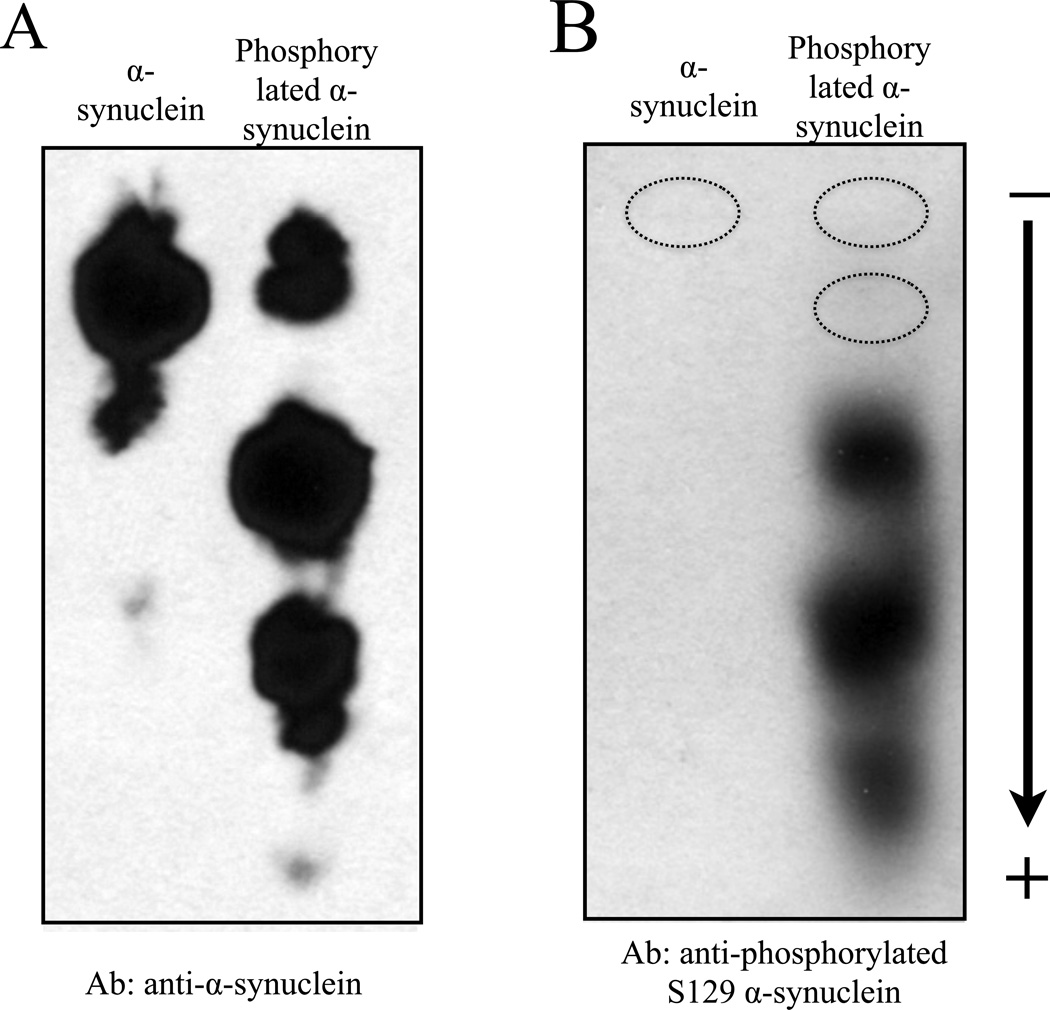
Figure 2. Immunoblotting application following NUT-PAGE.
Following 10% NUT-PAGE resolution, unmodified and phosphorylated α-synuclein were transferred to a nitrocellulose membrane, and probed with a general anti-α-synuclein antibody (A) or anti-phosphorylated Ser129 antibody (B) using standard western blotting procedures. Note that panels A and B were from two independent protein preps and PAGE runs. A parallel anti-α-synuclein immunoblot of that shown in panel B allowed us to mark the bands not detectable by the phosphorylation-specific antibody (dotted circles), suggesting that Ser129 is phosphorylated only in the presence of other pre-existing phosphorylation events.

