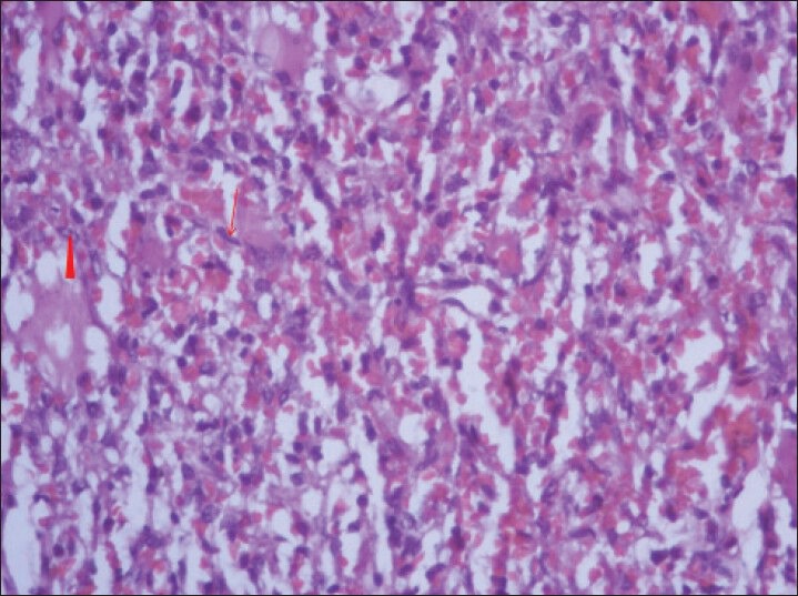
Figure 11.

40-year-old female with abdominal mass subsequently diagnosed as EGIST in retroperitoneum. Photomicrograph of hematoxylin and eosin (H and E) stained sample (×40) shows fascicles of uniform bland spindle cells (thin long red arrow) with pale eosinophilic fibrillary cytoplasm and extravasated red blood cells. Perinuclear vacuoles indent the nucleus (red arrowhead).
