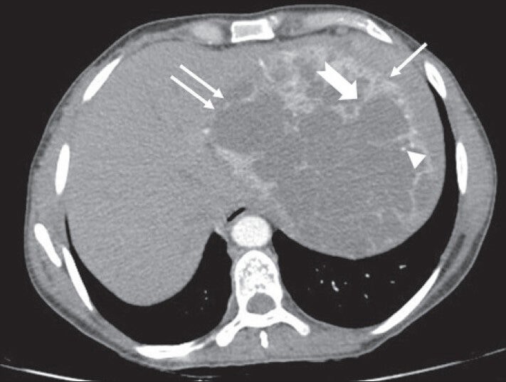
Figure 3.

40-year-old female with abdominal mass subsequently diagnosed as EGIST in the retroperitoneum. CECT abdomen (axial section) during venous phase shows the lesion compressing spleen (solid arrow), left lobe of liver (solid double arrows) without invasion (negative embedded organ sign). Few septae in the periphery appear thicker (notched arrow). Few tiny foci of calcification are also noted in the periphery (arrowhead)
