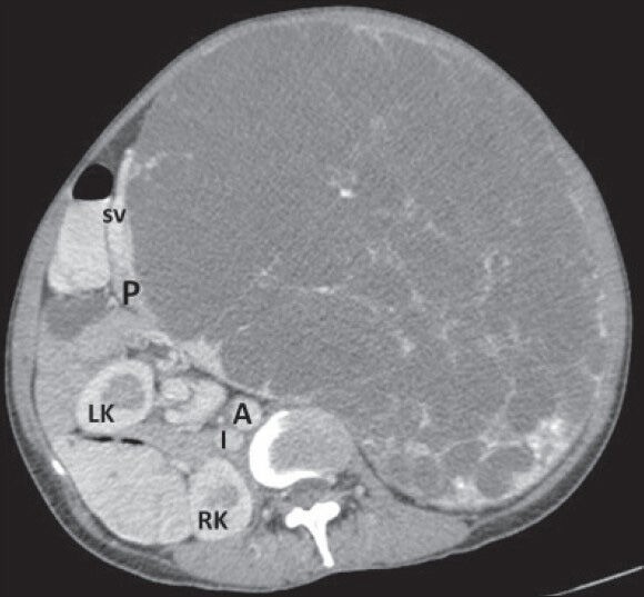
Figure 4.

40-year-old female with abdominal mass subsequently diagnosed as EGIST in retroperitoneum. CECT abdomen (axial section) during venous phase shows the lesion compressing head and body of pancreas (P) without infiltration (negative embedded organ sign) and a distinct intervening fat plane with abdominal aorta (A). LK: Left kidney, RK: Right kidney, I: Inferior vena cava, SV: Splenic vein.
