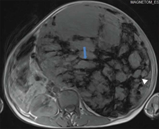
Figure 7.

40-year-old female with abdominal mass subsequently diagnosed as EGIST in retroperitoneum. Non-enhanced magnetic resonance imaging (MRI) (axial section) of abdomen shows the mass with low signal intensity areas, multiple markedly hypointense linear bands (blue arrow) in a reticular pattern, and few hypointense round foci (white arrowhead) on T1-weighted spin echo (SE) images.
