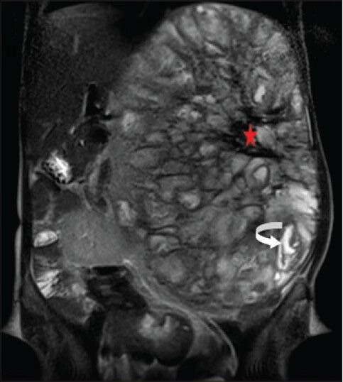
Figure 9.

40-year-old female with abdominal mass subsequently diagnosed as EGIST in retroperitoneum. MRI (coronal section) of abdomen shows a few of the loculated structures with bright fluid signal intensity (curved white arrow) and the central region of the lesion with large hypointense signal (red asterisk) on T2-weighted FSE images.
