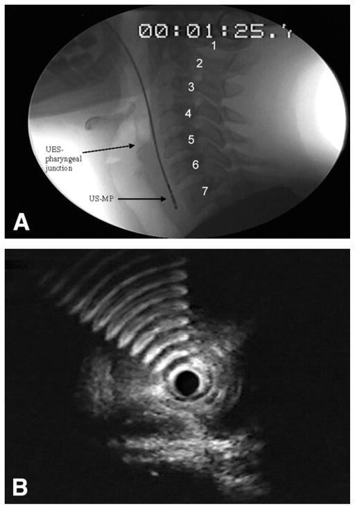
Figure 1.
A, Representative fluoroscopic image of cervical vertebrae (1–7) used as a stationary point of reference for placing 0.5-cm intervals above and below the UES-pharyngeal junction. UES, upper esophageal sphincter; US-MP, US miniprobe. B, Hyperechoic artifacts abruptly appear when the transducer of the US-MP is positioned at the level of the UES-pharyngeal junction during station pull through. This image demonstrates the air-tissue interface where air artifacts are first noted anterior to the flat-shaped muscle.

