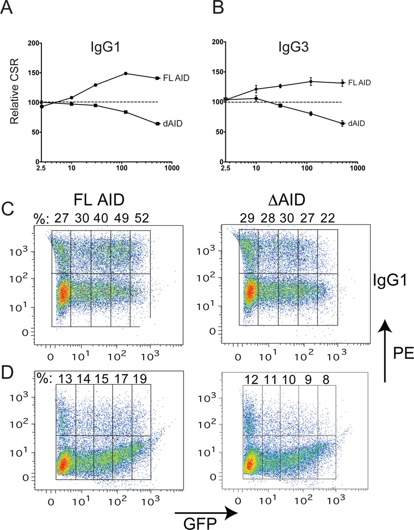
Figure 2. Dose-response assays of IgG1 (A, C) and IgG3 (B, D) switching in aid+/+ mouse splenic B cells expressing increasing levels of AID-ER or ΔAID-ER, as assayed by GFP expression (MFI).
Each data point on the curves in A and B represent the average%IgG1 or %IgG3 (±SEM) at the indicated MFI (binned as shown in C and D) of 3 cultures from 1 mouse transduced with the indicated retrovirus relative to %CSR in cells transduced with the empty ER retrovirus. Data points on the Y-axis (MFI=2.5) in A and B represent CSR in GFP-negative cells, expressing only endogenous AID. CSR in the GFP-negative cells are shown in the left most boxes of the FACS dot plots in C and D. The procedure for gating is illustrated in Fig 7A.

