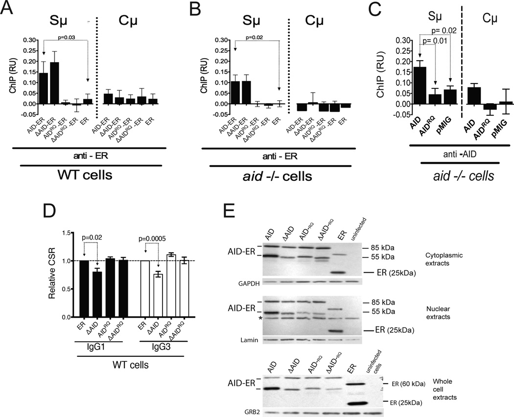
Figure 5. Both the DN effect and association of ΔAID-ER with Sµ depend upon deaminase activity of ΔAID-ER.
(A) ChIP of the AID-ER proteins at Sµ and Cµ in WT cells relative to input DNA, using anti-ER antibody. Error bars indicate SEM. 5 ChIPs (2 independent experiments, 2 mice) were performed, except for the no antibody control for FL-AID, where 4 were performed. ChIPs were analyzed by qPCR; % input was calculated, and % input in absence of antibody was subtracted. (B) ChIP of the AID-ER proteins at Sµ and Cµ in aid−/− cells relative to input DNA. 6 ChIPs for AID, ΔAID, and ER and 3 ChIPs for AIDRQ and ΔAIDRQ were performed (2 mice). Analysis as in A. (C) ChIP experiments performed with aid−/− cells transduced with untagged AID pMIG constructs, immunoprecipitated with anti-AID antibody (10). Error bars indicate SEM. 5–6 ChIPs (2 independent experiments, 2 mice) were performed. ChIPs were analyzed by qPCR; % input was calculated, and % input in absence of antibody was subtracted. P values determined by the two-tailed T test. (D) Compilation of IgG1 and IgG3 CSR results (+SEM) for the indicated RV-AID constructs in GFP-hi WT splenic B cells. Results are normalized to AID-ER results in one of the cultures each for IgG1 and IgG3. Six cultures (3 mice, 2 cultures each) were performed. CSR is plotted for GFP-hi cells, which are ~50% of the total GFP+ cells. Cells transduced with ER switch similarly to GFP-negative cells in the same cultures (not depicted). (E) Western blots of AID-ER in nuclear, cytoplasmic, and whole cell extracts from transduced WT splenic B cells. 20 µg protein was loaded in each lane. *Unknown irrelevant protein.

