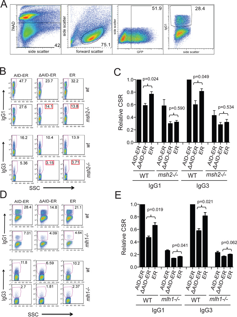
Figure 7. FACS analyses of CSR in WT, msh2−/−, and mlh1−/− cells demonstrates that the DN effect of ΔAID-ER requires the presence of Msh2.
(A) Gating strategy used for all FACS experiments in this manuscript, proceeding from left to right. Example is from panel D: WT cells transduced with AID-ER transduced and induced to switch to IgG1. Retrovirally-infected cultured cells were stained with 7-AAD to detect dying/dead cells, and with antibody (Fab’2) to IgG1 or IgG3 conjugated to PE. The percent of viable (7AAD-neg), retrovirally-infected (GFP+) cells that were IgG1+ of IgG3+ was determined as shown. Compensation and gating were performed using FlowJo software (Treestar). (B) FACS results for one representative CSR experiment comparing the DN effect of ΔAID-ER in WT and msh2−/− B cells (expressing endogenous AID). The FACS plots show only viable and GFP+ cells; the gates represent PE-IgG1/PE-IgG3 positive cells within the GFP+ populations as indicated. The entire GFP+ population was analyzed in these experiments. Side scatter (SSC) is plotted on the X-axis. (C) Compilation of IgG1 and IgG3 CSR results (+SEM) for the indicated constructs in WT and msh2−/− cells relative to CSR in WT cells expressing AID-ER. Two independent experiments (2 mice, 3 cultures each) were performed. IgG1 and IgG3 CSR are significantly increased in WT cells expressing AID-ER relative to ER (p=0.004 and 0.011, respectively). IgG1 and IgG3 CSR in msh2−/− cells expressing AID-ER relative to ER were not significantly increased (p=0.065 and 0.284, respectively). (D) FACS results for one experiment comparing the DN effect of ΔAID-ER in WT and mlh1−/− B cells. (E) Compilation of IgG1 and IgG3 CSR results (+SEM) for the indicated constructs in WT and mlh1−/− cells relative to CSR in WT cells expressing AID-ER). Three independent experiments (3 mice, 3 cultures each) were performed. IgG1 and IgG3 CSR were significantly increased in WT cells expressing AID-ER relative to ER (p=0.002 and 0.027, respectively). IgG1 CSR in mlh1−/− cells expressing AID-ER relative to ER was significantly increased (p=0.004) but not IgG3 CSR (p=0.256).

