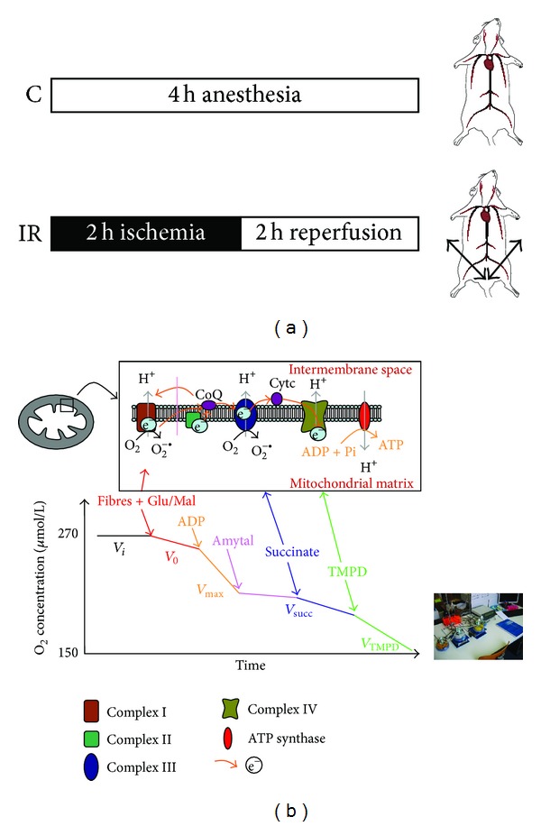
Figure 1.

(a) Experimental design. C: control; IR: ischemia-reperfusion. (b) Schematic representation of the mitochondrial respiratory chain with specific substrates and inhibitors. CI: complex I (NADH-CoQ reductase), CII: complex II (succinate-CoQ reductase), CIII: complex III (CoQH2-c reductase), CIV: complex IV (cytochrome c oxidase, COX), and TMPD: N, N, N′, N′-tetramethyl-p-phenylenediamine dihydrochloride. Schematic oxygraph trace showing oxygen consumption by the permeabilized skeletal myofibers, using indicated substrates and inhibitors: V 0, before ADP; V max, complexes I, III, and IV activities, using glutamate and malate; V succ, complexes II, III, and IV activities, using succinate; V TMPD/asc, complex IV activity using TMPD/Ascorbate.
