
Figure 1.
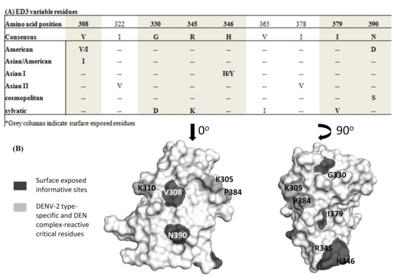
ED3 of different DENV-2 genotypes. Panel A: Alignment representing the ED3 informative sites, the residues that are bolded are surface exposed. Panel B: Structure of ED3 with the type-specific and complex-reactive critical residues highlighted light gray, and the surface exposed informative sites highlighted in dark gray.