
FIGURE 4:
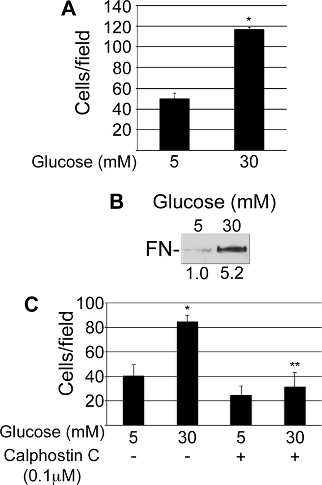
High glucose induces activation of β1 integrins. Mesangial cells were grown for 3 d in normal or high glucose (A, B) or for 2 d in normal or high glucose with dimethyl sulfoxide or 0.1 μM calphostin C (C). (A, C) Cells were then plated on surfaces coated with 9EG7 anti-β1 antibody and incubated for 30 min at 37°C before unattached cells were washed away. Attached cells were fixed, and three wells/condition were counted. Bar graph shows the mean of two independent experiments ± 1 SD; *p < 0.05 compared with 5 mM glucose; **p < 0.05 compared with 30 mM glucose. (B) Cells in suspension were incubated with rat FN for 30 min at room temperature, pelleted through a sucrose cushion, lysed in SDS sample buffer, and immunoblotted with IC3 anti-rat FN monoclonal antibody. Relative densitometry values (below the lanes) are the mean of three experiments.